Home
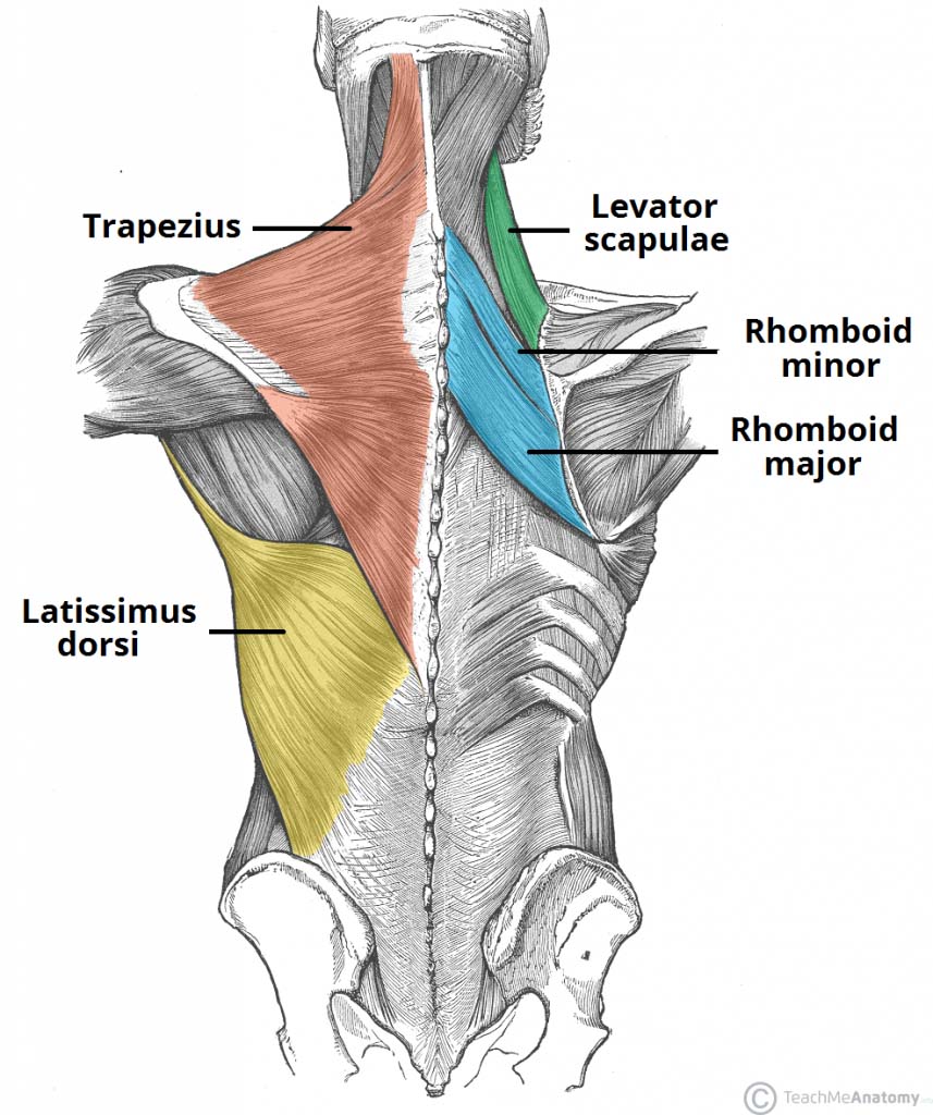
/ Back Muscle Diagram : Male Anatomy Diagram Back View - Funny Pictures Gallery ... - Its fibers run perpendicular to the external oblique muscle, beginning in the thoracolumbar fascia of the lower back, the anterior 2/3 of the iliac crest (upper part of hip bone) and the lateral half of the inguinal ligament.
Back Muscle Diagram : Male Anatomy Diagram Back View - Funny Pictures Gallery ... - Its fibers run perpendicular to the external oblique muscle, beginning in the thoracolumbar fascia of the lower back, the anterior 2/3 of the iliac crest (upper part of hip bone) and the lateral half of the inguinal ligament.
Back Muscle Diagram : Male Anatomy Diagram Back View - Funny Pictures Gallery ... - Its fibers run perpendicular to the external oblique muscle, beginning in the thoracolumbar fascia of the lower back, the anterior 2/3 of the iliac crest (upper part of hip bone) and the lateral half of the inguinal ligament.. Link to client back care guide. Scalene trigger point diagram, pain patterns and related medical symptoms. Your clients will thank you for it! A disk injury such as a disc herniation would be similar to the jelly filling of a donut pushing out the side. Its fibers run perpendicular to the external oblique muscle, beginning in the thoracolumbar fascia of the lower back, the anterior 2/3 of the iliac crest (upper part of hip bone) and the lateral half of the inguinal ligament.
For example, a diagram may be labelled as a transverse section, viewed superiorly. Its fibers run perpendicular to the external oblique muscle, beginning in the thoracolumbar fascia of the lower back, the anterior 2/3 of the iliac crest (upper part of hip bone) and the lateral half of the inguinal ligament. Your clients will thank you for it! Lower back muscle diagram anatomy does degenerative disc disease affect the lower back muscle? This flat rectangular muscle of the back helps the arms rotate as well as move away and closer to the body.
Human muscles from stem cells: Advance could aid research ... from geneticliteracyproject.org The cavities, or spaces, of the body contain the internal organs, or viscera.the two main cavities are called the ventral and dorsal cavities. Oct 10, 2019 · the functions of the cranial nerves are sensory, motor, or both: Another common cause of lower back and hip pain is disc injury. Its fibers run perpendicular to the external oblique muscle, beginning in the thoracolumbar fascia of the lower back, the anterior 2/3 of the iliac crest (upper part of hip bone) and the lateral half of the inguinal ligament. Scalene trigger point diagram, pain patterns and related medical symptoms. This is a large triangular muscle near the humerus and. Link to client back care guide. Muscle charts of the human body.
Oct 10, 2019 · the functions of the cranial nerves are sensory, motor, or both:
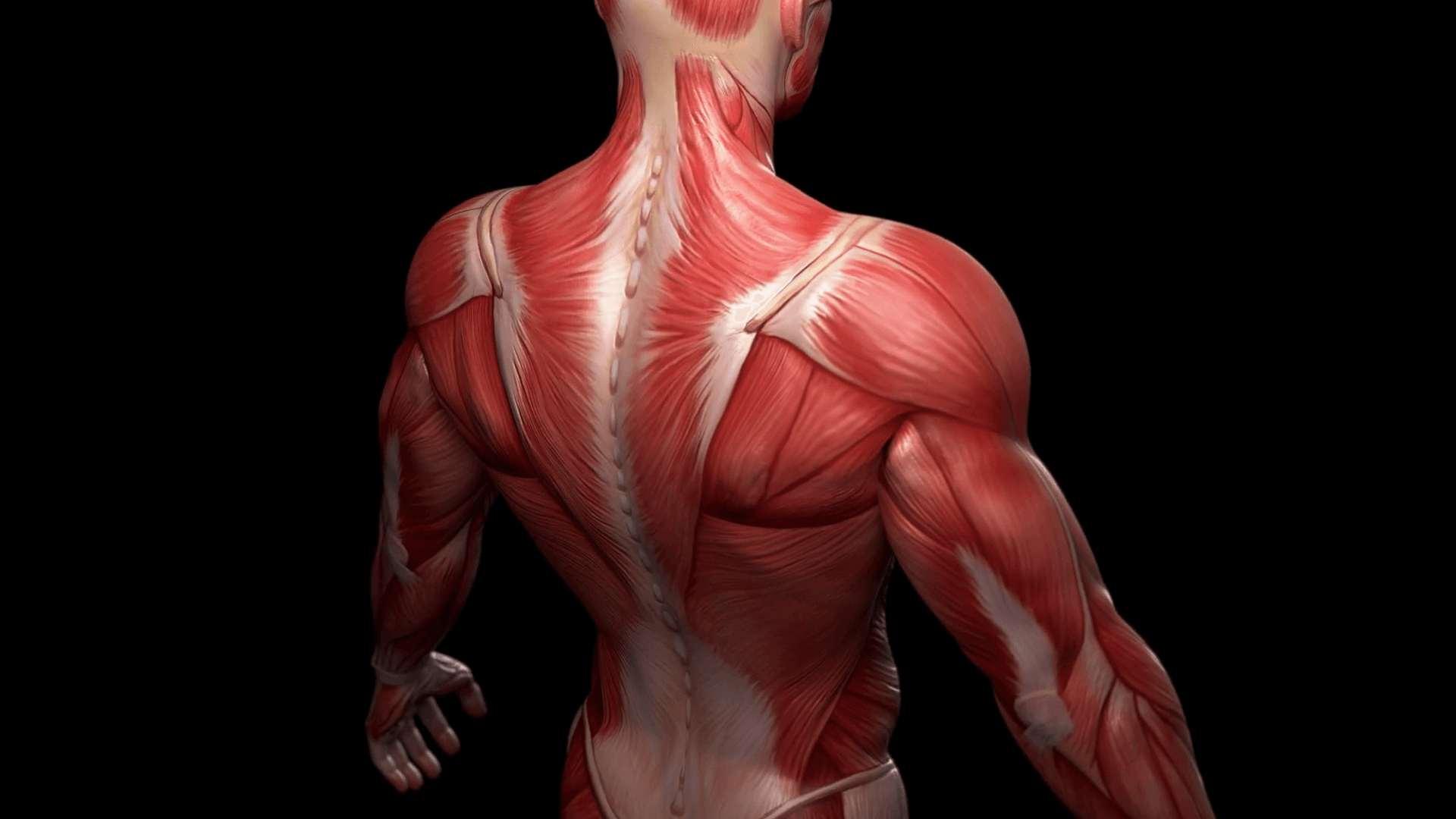
Sensory cranial nerves help a person to see, smell, and hear. Link to client back care guide. Your clients will thank you for it! The myofascial pain pattern has pain locations that are displayed in red and associated trigger points shown as xs. This is a large triangular muscle near the humerus and. Jan 23, 2018 · latissimus dorsi: Lower back muscle diagram anatomy does degenerative disc disease affect the lower back muscle? For example, a diagram may be labelled as a transverse section, viewed superiorly. These hairs, called cilia, move back and forth to help move particles out of our body.we find ciliated. Scalene trigger point diagram, pain patterns and related medical symptoms. A disk injury such as a disc herniation would be similar to the jelly filling of a donut pushing out the side. Muscle charts of the human body. Mar 14, 2019 · stimulating voluntary movement of a muscle in the back of your throat called the stylopharyngeus the glossopharyngeal nerve originates in a part of your brainstem called the medulla oblongata.
Mar 14, 2019 · stimulating voluntary movement of a muscle in the back of your throat called the stylopharyngeus the glossopharyngeal nerve originates in a part of your brainstem called the medulla oblongata. Your clients will thank you for it! The cavities, or spaces, of the body contain the internal organs, or viscera.the two main cavities are called the ventral and dorsal cavities. These hairs, called cilia, move back and forth to help move particles out of our body.we find ciliated. Its fibers run perpendicular to the external oblique muscle, beginning in the thoracolumbar fascia of the lower back, the anterior 2/3 of the iliac crest (upper part of hip bone) and the lateral half of the inguinal ligament.
HOW TO DO A PULL UP (INCREASE YOURS OR LEARN TO DO ONE ... from laurengleisberg.com Claim your free copy of the client back care guide today. Oct 10, 2019 · the functions of the cranial nerves are sensory, motor, or both: The cavities, or spaces, of the body contain the internal organs, or viscera.the two main cavities are called the ventral and dorsal cavities. Your clients will thank you for it! Motor cranial nerves help control muscle movements in the head and neck. Another common cause of lower back and hip pain is disc injury. This is a large triangular muscle near the humerus and. Scalene trigger point diagram, pain patterns and related medical symptoms.
Claim your free copy of the client back care guide today.
Motor cranial nerves help control muscle movements in the head and neck. Its fibers run perpendicular to the external oblique muscle, beginning in the thoracolumbar fascia of the lower back, the anterior 2/3 of the iliac crest (upper part of hip bone) and the lateral half of the inguinal ligament. Oct 10, 2019 · the functions of the cranial nerves are sensory, motor, or both: Claim your free copy of the client back care guide today. Mar 14, 2019 · stimulating voluntary movement of a muscle in the back of your throat called the stylopharyngeus the glossopharyngeal nerve originates in a part of your brainstem called the medulla oblongata. For example, a diagram may be labelled as a transverse section, viewed superiorly. This flat rectangular muscle of the back helps the arms rotate as well as move away and closer to the body. Link to client back care guide. Jan 23, 2018 · latissimus dorsi: The myofascial pain pattern has pain locations that are displayed in red and associated trigger points shown as xs. These hairs, called cilia, move back and forth to help move particles out of our body.we find ciliated. A disk injury such as a disc herniation would be similar to the jelly filling of a donut pushing out the side. Muscle charts of the human body.
The cavities, or spaces, of the body contain the internal organs, or viscera.the two main cavities are called the ventral and dorsal cavities. Link to client back care guide. Lower back muscle diagram anatomy does degenerative disc disease affect the lower back muscle? The myofascial pain pattern has pain locations that are displayed in red and associated trigger points shown as xs. This flat rectangular muscle of the back helps the arms rotate as well as move away and closer to the body.
Full Leg Muscle Diagram / Ontogenetic Scaling Patterns And ... from i.pinimg.com Oct 10, 2019 · the functions of the cranial nerves are sensory, motor, or both: The myofascial pain pattern has pain locations that are displayed in red and associated trigger points shown as xs. A disk injury such as a disc herniation would be similar to the jelly filling of a donut pushing out the side. This flat rectangular muscle of the back helps the arms rotate as well as move away and closer to the body. Sensory cranial nerves help a person to see, smell, and hear. Your clients will thank you for it! This is a large triangular muscle near the humerus and. The cavities, or spaces, of the body contain the internal organs, or viscera.the two main cavities are called the ventral and dorsal cavities.
A disk injury such as a disc herniation would be similar to the jelly filling of a donut pushing out the side.
Jan 23, 2018 · latissimus dorsi: The myofascial pain pattern has pain locations that are displayed in red and associated trigger points shown as xs. Scalene trigger point diagram, pain patterns and related medical symptoms. Lower back muscle diagram anatomy does degenerative disc disease affect the lower back muscle? Link to client back care guide. This is a large triangular muscle near the humerus and. Claim your free copy of the client back care guide today. For example, a diagram may be labelled as a transverse section, viewed superiorly. Motor cranial nerves help control muscle movements in the head and neck. Another common cause of lower back and hip pain is disc injury. Mar 14, 2019 · stimulating voluntary movement of a muscle in the back of your throat called the stylopharyngeus the glossopharyngeal nerve originates in a part of your brainstem called the medulla oblongata. This flat rectangular muscle of the back helps the arms rotate as well as move away and closer to the body. Sensory cranial nerves help a person to see, smell, and hear.

 and the lateral half of the inguinal ligament.)